The Science of Suspended Animation in Killifish


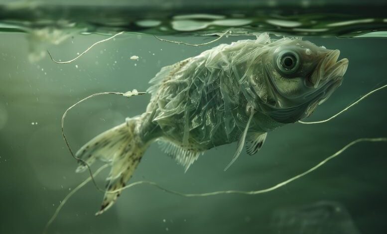
African turquoise killifish embryos survive dry seasons by entering diapause, using ancient genes to halt and resume embryonic development. Similar genetic expressions are found in other species, including mice. (Artist’s concept of a fish, rather than an embryo, in suspended animation.) Credit: SciTechDaily.com
How Killifish Embryos Use Suspended Animation To Survive Over 8 Months of Drought
The African turquoise killifish utilizes a unique survival strategy through diapause, leveraging ancient genes to endure dry seasons. This mechanism involves pausing embryonic development, which is later resumed. Research also indicates similar genetic patterns in other species, suggesting a shared evolutionary trait for suspended animation.
Suspended Animation in African Turquoise Killifish
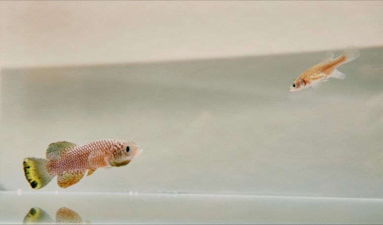
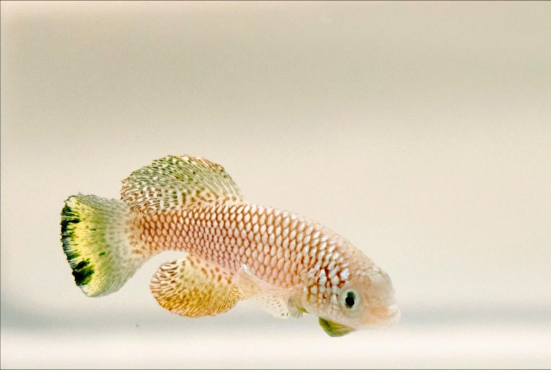
Transient ponds in Zimbabwe and Mozambique are home to African turquoise killifish. To survive the annual dry season, the fish’s embryos enter a state of extreme suspended animation or “diapause” for approximately 8 months.
A new study has revealed the evolutionary adaptations that allow the killifish to sustain this remarkable survival technique. Published on May 30 in the journal Cell, the findings indicate that killifish developed the ability to diapause less than 18 million years ago by co-opting ancient genes that originated more than 473 million years ago. Comparative analyses further demonstrate that similar gene expression patterns during diapause are found in other animals, including the house mouse.
“The whole program is like day and night—there is life in the normal state and life in the diapause state, and the way this happened was by reshuffling or re-wiring the regulatory region of a whole set of genes,” says senior author and molecular biologist Anne Brunet of Stanford University.
Rapid Maturation and Reproduction of Killifish
African turquoise killifish mature faster than any other vertebrate species, and adults live for only around 6 months, even in captivity. The fish reproduce rapidly before their watery homes disappear, but their embryos remain behind in the dry mud, ready to hatch when the next year’s rains come. Embryonic diapause also occurs in other vertebrate species, including fish, reptiles, and some mammals, but killifish diapause is remarkably extreme because it lasts for such an extended period (8 months on average and up to 2 years in the lab) and because killifish embryos enter suspended animation much later in development than other animals.
“It’s roughly in the middle of development, and many organs are already formed by that stage— they have a developing brain and a heart which stops beating in diapause and then starts again,” says first author Param Priya Singh of the University of California, San Francisco. “Killifish are the only vertebrate species that we know of that can undergo diapause so late in development.”
Detailed Gene Analysis During Diapause
To understand diapause evolution, the research team first characterized the gene expression of the African turquoise killifish (Nothobranchius furzeri) during different developmental stages. They focused on duplicated copies of genes called “paralogs,” because gene duplication is one of the primary mechanisms by which new genes originate and specialize. Overall, the researchers identified 6,247 paralog pairs that exhibited specialized gene expression patterns during diapause. Surprisingly, they estimated that most of the diapause-specialized genes were “very ancient” paralogs, having originated more than 473 million years ago.
“Even though diapause evolved relatively recently, the genes that are specialized in diapause are really ancient,” said Brunet. “We found that most of the genes that specialize for diapause in killifish are very ancient paralogs, which means that they were duplicated in the common ancestor of all vertebrates.”
Comparative Analysis Across Species
Since diapause also occurs in some other species of killifish, the researchers compared gene expression between embryos of the African turquoise killifish, the South American killifish (Austrofundulus limnaeus), which also undergoes diapause, and two killifish species that do not undergo diapause, the red-striped killifish (Aphyosemion striatum) and lyretail killifish (Aphyosemion austral).
They found significant overlap in gene expression patterns between the African turquoise and South American killifish, which evolved diapause independently of each other, but not in the two non-diapausing species. Likewise, the researchers found significant correlation in the gene expression patterns of house mouse (Mus musculus) embryos during diapause and showed that diapause-specialized genes in mice also have very ancient origins.
“This suggests that the same mechanisms that enable diapause have been repeatedly co-opted for the evolution of diapause across distantly related species,” says Singh.
Next, the investigators explored how these diapause-specialized genes are regulated in the killifish. They identified several key transcription factors that control the altered gene expression patterns seen during diapause, including REST and FOXO3, which are known to be expressed during hibernation (a different form of suspended animation) in mammals. Notably, several of these regulatory genes are involved in lipid metabolism, which has a distinctive profile during diapause.
“One of the key elements of diapause is this special lipid metabolism,” said Brunet. “During diapause, they seem to have much higher levels of triglycerides and very long chain fatty acids, which are forms of storage and also perhaps aid with long-term protection of the organism’s membranes.”
Future Directions in Diapause Research
The team plans to continue investigating how different species regulate diapause and dig deeper into the role of lipid metabolism during diapause and other types of suspended animation.
“It’s such a complex state that I think we are just scratching the surface,” said Singh. “We want to go deeper into specific aspects of how lipid metabolism is regulated during diapause, and we are also interested in examining the role of specific cell types during diapause.”
Reference: “Evolution of diapause in the African turquoise killifish by remodeling the ancient gene regulatory landscape” by Param Priya Singh, G. Adam Reeves, Kévin Contrepois, Katharina Papsdorf, Jason W. Miklas, Mathew Ellenberger, Chi-Kuo Hu, Michael P. Snyder and Anne Brunet, 28 May 2024, Cell.
DOI: 10.1016/j.cell.2024.04.048
This research was supported by the Glenn Foundation for Medical Research, the NOMIS Distinguished Scientist and Scholar award, the Stanford Center for Computational, Evolutionary, and Human Genomics, the National Institutes of Health, and the National Science Foundation

